Specific Viral Families: Retroviridae


Retroviridae

The retroviruses are enveloped viruses that have two complete copies of (+) sense RNA. They also contain the enzyme reverse transcriptase which uses the viral RNA to form a complementary strand of DNA, which is then replicated to form a dsDNA. For the genetic information to be transcribed and translated, this DNA migrates to the host cell nucleus and become incorporated into chromosomes of host cells. Such integrated viral DNA is known as a provirus.

http://pathmicro.med.sc.edu/lecture/RNA2.jpg

Retroviruses cause tumours and leukemia in humans. The human retroviruses invade immune defense cells called T lymphocytes and are referred to as Human T cell Leukemia Viruses (HTLV). Both HTLV-1 and HTLV-2 are associated with malignancies (leukemia and other tumours), whereas the human immunodeficiency virus (HIV) causes acquired immune deficiency syndrome (AIDS).

Important Properties

    · Spherical enveloped virion, 80-100nm

    · Ribonucleoprotein in central nucleoid (concentric or truncated cone in lentivurses) within icosahedral capsid.

    · Envelope with glycoprotein peplomers

    · 2 copies of linear plus sense ssRNA each 7-10kb

    · 3’ polyadenylated tail and 5’ cap

    · Reverse transcriptase

    · Formation of long terminal repeats before prpovirus DNA inserted into host genome

    · Genome consists of gag, pol, env genes some regulatory genes, some oncogenes.

Human Immunodeficiency Virus

HIV is one of the human T-cell lymphotropic retroviruses. HIV preferentially infects and kills helper (CD4) T lymphocytes, resulting in a loss of cell mediated immunity and a high probability that the host will develop opportunistic infections. Other cells (e.g. macrophages and monocytes) that have CD4 proteins on their surfaces can be infected also.

https://blogger.googleusercontent.com/img/b/R29vZ2xl/AVvXsEhuSD_M4_xi0Hr1WcOR7MVcKfGUBOjidoN8TQMWhyphenhyphencbizC-ehgadoZnvoMqDJ1b4rauaIlZmedd4GIWq17Ah6UgpiqOVe9-wHK3nVKkZiFZfZhBUp89-23d0kfHIY7_oxrc0nZvkCsDLVpQ/s1600-h/aaaaad.jpg

Genes and Proteins of the Human Immunodeficiency Virus

Gene

Proteins Encoded by Gene

Function of Proteins

I. Structural Genes Found in All Retroviruses

gag

p24, p7

p17

Nucleocapsid

Matrix

pol

Reverse Transciptase

Protease

Integrase

Transcribes RNA genome into DNA

Cleaves precursor polypeptide

Integrates viral DNA into host cell DNA

env

gp120

gp41

Attachment to CD4 protein

Fusion with host cell

II. Regulatory Genes Found in Human Immunodeficiency Virus that are Required for Replication

tat

Tat

Activation of transcription of viral genes

rev

Rev

Transport of late mRNAs from nucleus to cytoplasm

III. Regulatory Genes Found in Human Immunodeficiency Virus that are NOT Required for Replication (accessory genes)

nef

Nef

Decreases CD4 proteins and class I MHC proteins on surface of infected cells; induces death of uninfected cytotoxic T cells; important for pathogenesis by SIV

vif

Vif

Stabilizes newly synthesized viral DNA and transports core through cytoplasm

vpr

Vpr

Transports viral core from cytoplasm into nucleus in non-dividng cells

vpu

Vpu

Enhances virion release from cell

Replicative Cycle of HIV

The replication of HIV follows the typical retroviral cycle.

  1. Inital step in the entry of HIV into the cell is the binding of the virion gp120 envelope protein to the CD4 protein on the cell surface.

  2. The virion gp120 protein then interacts with a second protein on the cell surface, one of the chemokine receptors (CXCR4 and CCR5). Individuals with mutations in the gene encoding CCR5 have protection from infection with HIV.

  3. The virion gp42 protein mediates fusion of the viral envelope with the cell membrane, and the virion enters the cell.

  4. After uncoating, the virion RNA-dependent DNA polymerase transcribes the genome RNA into double-stranded DNA, which integrates into the host cell DNA, mediated by integrase.

  5. The viral DNA can integrate at different sites in the host cell DNA, and multiple copies of viral DNA can integrate.

  6. Viral mRNA is transcribed from the proviral DNA by host cell ENA polymerase and translated into several large polyproteins.

  7. Precursor polyproteins can be assembled into nucleocapsid.

  8. As immature virions buds from the cell membrane, the polyproteins are cleaved by the viral protease. This results in the formation of mature, infectious virions.


http://www.nature.com/embor/journal/v4/n6s/images/embor857-f2.gif

Transmission

    · Transfer of infected blood.

    · Sexual contact (horizontal transfer)

    · Perinatal transmission from infected mother to neonate: Either across the placenta, at birth, or via breast milk. (vertical transfer)

Pathogenesis

http://www.nature.com/nri/journal/v3/n4/images/nri1058-f3.gif

  · Primary Infection

o Acute stage

o Flu-like symptoms

o Fever

o Skin rash

o Swollen skin nodes

o Due to virulence factors such as rate of replication, propensity to mutate and cytopathogenicity.

o Also due to host resistance mechanisms such as the suppression by CD8 T suppressor cells and the presence of cytotoxic T-lymphocytes.

    · Asymptomatic Stage

o No apparent disease

o Fall in CD4 T lymphocytes

o Possible signs:

§ Fatigue

§ Depression

§ Weight loss

§ Memory disorders

    · Symptomatic Stage

o AIDS-related complex

§ Diseases not considered definitive of AIDS

§ May be attributed to HIV infection

§ Indicative of defect in cell-mediate immunity

o AIDS

§ Opportunistic infections as a result of fall in CD4 T lymphocytes. (e.g. Kaposi’s Sarcoma)


http://www.aegis.com/images/aids-ois1.gif

Treatment

    · Highly active antiretroviral therapy (HAART)

o Consists of two nucleoside inhibitors (zidovudine and lamivudine) and a protease inhibitor (indinavir)

o Effective in prolonging life, improving the quality of life, and reducing viral load but does not cure the chronic HIV infection i.e. a latent infection of CD4+ cells continues indefinitely.

o Such combination therapy able to combat resistance—which arise due to rapid mutations due to inefficiency of reverse transcriptase.

    · Non-specific therapeutic management

    · Immunomodulation

o Enhancement of immune system through treatment with interleukin-2 (still under study)

    · No vaccine for human use is available.

Video from youtube: embed:

Prevention

Taking measures to avoid exposure to the virus:

    · Using condoms

    · Not sharing needles

    · Discarding donated blood that is contaminated with HIV

Specific Viral Families: Orthomyxoviridae


Orthomyxoviridae

The orthomyxoviruses are medium-sized, enveloped, (-)-sense RNA viruses that vary in shape from spherical to helical. Their genome is segmented into eight pieces. Orthomyxoviruses have an affinity for mucus. The influenza A and B viruses in this viral family are of particular medical importance to humans since they cause disease in humans.

Influenza Viruses

The influenza A virus is an orthomyxovirus that infects humans, birds, swine and horses. It causes pandemics of influenza (~10-20 years) and major outbreaks of influenza (virtually every year in various countries). The influenza B virus, on the other hand, appears to be specific to humans only. It causes major outbreaks of influenza, which occur less often as those caused by the influenza A virus.

http://www.ifpma.org/Influenza/content/images/diagram_virus.jpg

Diagram of the Influenza virus

Important Properties

    · Typically spherical (100nm in diameter).

    · Composed of a single stranded (-) sense RNA genome in eight segments, a helical nucleocapsid, and an outer lipoprotein envelope.

    · 3 polymerase polypeptides with each segment.

    · 5’ and 3’ end of all segments are highly conserved.

    · Virion contains RNA-dependent RNA polymerase, which transcribes the (-) sense RNA to mRNA. Thus, the genome is not infectious.

    · Viral envelope is covered with two different types of spikes—haemagglutinin (HA) and neuraminidase (NA).

    · The ratio of HA:NA is 5:1.

Haemagglutinin functions at the beginning of infection, whereas the neuraminidase functions at the end.

Haemagglutinin

    · Binds to the cell surface receptor (neuraminic acid, sialic acid) to initiate infection.

    · Target of neutralizing antibody.

Neuraminidase

    · Cleaves neuraminic acid (sialic acid) to release progeny virus from the infected cell.

    · Degrades the protective layer of mucus in the respiratory tract, thus enhancing the ability of the virus to infect the respiratory epithelium.

Pathogenesis

    · After the virus has been inhaled, the neuraminidase degrades the protective mucus layer, allowing the virus to gain access to the cells of the upper and lower respiratory tract.

    · The infection is limited primarily to this area because the HA receptors have a specific affinity for the epithelial cells of the respiratory tract.

    · Despite systemic symptoms, viremia rarely occurs.

    · The systemic symptoms are due to cytokines circulating in the blood.

    · There is necrosis of the superficial layers of the respiratory epithelium.

    · Immunity rests mainly upon secretory IgA in the respiratory tract.

    · IgG is also produced and confers protection against future infections.

    · Innate resistance also plays a role in immunity—a mucus blanket and cilia helps to trap and expel the influenza virus out of the respiratory tract.

    · Cytotoxic T cells also play a protective role.

    · Immunocompromised patients especially prone to secondary infection such as pneumonia.

http://www.influenzareport.com/ir/images/image27.jpg

Clinical Features

    · After an incubation period 24-48 hours, fever, myalgias, headache, sore throat, and cough develop suddenly.

    · Severe myalgias (muscle pains) coupled with respiratory tract symptoms are typical of influenza.

    · Vomiting and diarrhoea are rare.

    · Symptoms usually resolve spontaneously in 4-7 days.

Differentiating Between the Common Cold and Flu

Laboratory Diagnosis

    · Nasal/throat washings or swabs; sputum.

    · Virus culture in MDCK cell line or chick embryo.

    · Direct ELISA for Haemagglutinin

    · PCR assay

Transmission and Epidemiology

    · Virus is transmitted by airborne respiratory droplets.

    · Ability of influenza A virus to cause epidemics is dependent on antigenic changes in haemagglutinin and neuraminidase.

    · Influenza infections are found all year round.

    · In the northern hemisphere, influenza occurs primarily in the winter months.

    · In the southern hemisphere, influenza occurs primarily in the winter months of June through August.

    · Few serotypes circulating simultaneously.

    · Epidemics and pandemics occur when the antigenicity of the virus has changed sufficiently that the pre-existing immunity of many people is no longer effective.

    · The antigenicity of influenza B virus also varies but not as dramatically or as often.

Antigenic Drift vs. Antigenic Shift

Antigenic Drift

Antigenic Shift

Minor change in genome.

Major change in genome.

Occurs in both Influenza A and B viruses.

Occurs in only Influenza A virus.

Point mutation in the genetic code of surface antigens.

Gene reassortment, where entire segments of RNA are exchanged, each one of which codes for a single protein (e.g. haemagglutinin).

Results in a new strain.

Results in a new subtype.

Happens all the time.

Happens occasionally.

Responsible for epidemics.

Responsible for pandemics.

https://blogger.googleusercontent.com/img/b/R29vZ2xl/AVvXsEiHqTazCXYaCPS7IC_6LrvBUpxQD7rlkXoTLlVRH5hb8c9mTJJFJLa6qQVqFiE3UTo_NdM4ZfFb7EK5O5TmZoiXuJh8QYoS4fs0kaK6yw6TxzUIzffq6PoKdZQdQqKMPCCkJUj2Bg_7Fxiz/s1600-h/aaaa.jpg

Strain Naming Convention

Strains of influenza are named as follows:

(Influenza Type)/(Animal species (omitted if human)/(Place of Isolation)/(Number of Isolates)/(Year of First Isolation)(H and N subtypes)

Example: A/Texas/36/91/(H1N1)

Control and Treatment

    · The main mode of prevention is the vaccine, which consists of killed influenza A and B viruses. It is rendered useless by antigenic shift/drift—therefore, the vaccine is usually reformulated every year to contain the current antigenic strains.

    · Antiviral drugs are used to treat influenza. This includes receptor analogues, transcriptase inhibitors, reverse transcriptase inhibitors, protease inhibitors and neuraminidase inhibitors etc.

http://www.pyroenergen.com/articles/images/tamiflu.jpg

Specific Viral Families: Picornaviridae


Picornaviridae

Picornaviruses are named for their small (“pico” + “RNA” = picorna) size. Nevertheless, they sure do have a massive and diverse array of viruses – over 100 serotypes! These viruses can even be traced all the way back to Ancient Egyptian records of polio epidemics, but are still around and cause a menagerie of diseases today.

Unique features, Morphology and Genome

Ø    Picornaviruses contain positive sense, single-stranded RNA that is approximately 7-8 kilobases long. 

Ø    The genome is monopartite  and polyadenylated at the 3’ end, but has a VPg protein at the 5’ end in place of a cap. 

Ø    The viral RNA is infectious and replication takes place in the cytoplasm

Ø    The virus has an IRES (Internal Ribosomal Entry Site) which distinguishes it from many other RNA viruses. 

Ø    The virus is naked with an icosahedral capsid. 

Ø    The triangulation number is 3, while the capsid has four unique proteins: VP1, 2, 3, and 4.

Ø    The capsid is one of the smallest of all viruses with a diameter of only 27-30nm.

Ø    Translation and cleavage of viral polypeptides produces eleven distinct proteins.


One example of a Picornaviridae virus is Rhinovirus.


With as many as more than 100 serotypes, rhinoviruses are transmitted through the respiratory route and replicate in the nose (“rhino”).  The many serotypes are then divided into “major” and “minor” groups and all cause a similar syndrome – the “common cold.”  The large number of serotypes allows many rhinovirus infections to occur in one person over time, since immunity only develops for one serotype and each newly acquired rhinovirus causes a new “cold.”  About half of all colds can be attributed to rhinoviruses, particularly those that occur in the winter.

Pathogenesis includes an Upper Respiratory Tract infection with a short incubation period of 2 to 3 days. Production of the endogenous IFN (Interferon) will help battle the virus to protect the body. IgA (a type of immunoglobulin present in blood and body secretions which may aid in fighting infections) is then locally synthesized, though it will degrade with time. However, our serum IgG (memory cell of immune system) would confer lifetime immunity! 

And how would you know that you’ve been infected by rhinovirus?

 Symptoms include, a watery nasal discharge, congestion, sneezing and little or no fever. 

A lab diagnosis of this virus will include culturing the virus, nasal washings, EIA and PCR. 

This epidemic occurs practically throughout the year, albeit there are a few serotypes circulating simultaneously! This is most probably due to the fact that the virus is abundant in nasal discharge. 

How does one control this “common cold” then? 

Well, you could wash your hands regularly, avoid touching your eyes or nose. If you happen to sneeze into a tissue, be sure to discard it right after! You might also want to avoid people who have a cold! Or, if you are the one with the cold, do stay at home! This will ensure that other people would not be affected! 


Rhino Virus

Specific Viral Families: Herpesviridae


Herpesviridae


http://pathmicro.med.sc.edu/virol/herpes_simplex.jpg

Above: TEM of Herpes Simplex Virus

Herpesviruses are a leading cause of human viral disease, second only to influenza and cold viruses. They are relatively large, enveloped viruses with linear dsDNA. Herpesviruses are widely distributed in nature.

Important properties

    · Concentric virion with

o Inner core

o Icosahedral capsid

o Amorphous tegument

o Envelope ( glycoprotein)

    · Linear dsDNA

    · Three origin of replication (ORI)

http://pathmicro.med.sc.edu/mhunt/dna15.jpg

Source:

The herpesviruses are known for their ability to cause latent infections.

    · In cells infected with herpesviruses, the viral dsDNA can exist as a provirus.

    · Herpesviruses remain in host cells, usually neurons, for long periods and retain the ability to replicate.

    · For example, a child who has recovered from chickenpox (varicella) will still have the virus in a latent form.

    · Years or decades later, the virus may be reactivated as a result of stress and/or physical factors.

    · This adult disease, which is very painful, is called shingles (zoster).

    · 11 of more than 100 genes of the herpesvirus genome are known to be involved in latency.

Pathogenesis of Human Herpes Type 1 to 5

Human Herpes Type

Name

Target cell Type

Disease

Latency

Transmission

1

Herpes simplex-1 (HSV-1)

Mucoepithelia

Oral herpes, encephalitis

Neuron

Close contact

2

Herpes simplex-2 (HSV-2)

Mucoepithelia

Genital and neonatal herpes, meningoencephalitis

Neuron

Close contact usually sexual

3

Varicella Zoster virus (VZV)

Mucoepithelia

Chickenpox (varicella) and Zoster (shingles)

Neuron

Contact or respiratory route

4

Epstein-Barr Virus (EBV)

B lymphocyte, epithelia

Infectious mononucleosis and Burkitt’s lymphoma; linked to Hodgkin’s disease, B cell lymphomas and to nasopharyngeal cancer.

B lymphocytes

Saliva

5

Cytomegalovirus (CMV)

Epithelia, monocytes, lymphocytes

Acute febrile illness; infections in immunosuppressed patients, leading cause of birth defects.

Monocytes, lymphocytes and possibly others

Contact, blood transfusions, transplantation, congenital

Clinical Features

HSV-1: Typically, Herpes labialis—Cold scores; Blisters around the mouth which lasts for around 1 week or more. May also affect the eyes (keratoconjunctivitis).

http://pathmicro.med.sc.edu/virol/coldsore2.jpg

HSV-2: Genital herpes, which is characterized by blisters, burning sensation and discharge.

VZV: In varicella, fever and malaise occur. Lesions appear on the trunk and spreads to the head and extremities. This is dangerous in pregnant women and may affect the nervous system—Guillain Barre syndrome. In zoster, painful vesicles occur along the course of a sensory nerve of the head or trunk.

http://pathmicro.med.sc.edu/virol/chickenpox3.jpg

Laboratory Diagnosis

    · Virus culture

    · Enzyme-linked immunosorbent assays (ELISAs)

    · Blood test

    · Tzanck smear

    · PCR assay

Epidemiology

HSV-1: Almost 100% of the adult population due to kissing and close proximity.

HSV-2: Up to 20% of the U.S. population due to sexual contact.

VZV: Varicella is a highly contagious disease of childhood; more than 90% of people in the U.S. have antibody by age 10 years. Varicella occurs worldwide.

EBV: Almost 100% of the adult population due to kissing and close proximity.

Control

HSV-1, HSV-2 and EBV: Avoid kissing to prevent contact with vesicular lesions or ulcers, and refrain from risky sexual behaviour. For the Herpes Simplex Viruses, caesarean section is recommended for women who are at term and who have genital lesions or positive viral cultures.

VZV: Vaccination with live, attenuated VZV (e.g. Varivax), and avoiding infected people.

Specific Viral Families: Hepadnaviridae


Hepadnaviridae

    · Small, enveloped, dsDNA (partially ssDNA) viruses.

    · The name hepadna comes from the infection of the liver—hepatitis—by a DNA virus.

    · Hepadnaviruses can cause chronic liver infections in humans and other animals, including ducks. In humans the hepatitis B virus causes hepatitis B, which can progress to liver cancer.

Hepatitis B Virus


http://pathmicro.med.sc.edu/lecture/hepatitis.gif

Important properties

  · 42-nm enveloped virion

    · Icosahedral nucleocapsid core contains a partially double-stranded circular DNA genome.

    · Envelope contains a protein called the surface antigen (HBsAg).

    · Core antigen (HBcAg) and the e antigen (HBeAg) are both located in the nucleocapsid protein but are antigenically different.

    · Endogeneous DNA-dependent DNA polymerase within the core.

    · Use of overlapping reading frame (ORF).

    · RNA-dependent DNA synthesis during replicative cycle.

Pathogenesis

    · Acute or chronic liver infection depending on the age at infection.

    · 90% of neonates and 50% of young children become chronically infected.

    · Only about 5% to 10% of immune-competent adults infected with HBV develop chronic hepatitis B.

    · A chronic carrier is someone who has HBsAg persisting in their blood for at least 6 months.

    · A high rate of heptatocellular carcinoma occurs in chronic carriers.

Basic characteristics of Hepatitis B infection:

Incubation period

60-90 days*

Fatality rate

1%

Recovery Rate

90%

Rate of Chronic Infection

<10%**

*with a range of 45-120 days

**although this can approach 90% in babies infected with HBV at birth.


http://www.medscape.com/content/2004/00/47/14/471470/art-jvh471470.fig2.gif

Above: The natural history of Hepatitis B

Clinical Features

Acute

    · Loss of appetite, nausea, vomiting, fever, abdominal pain and jaundice

    · About 90% - 95% of adults recover without sequelae

    · 5% - 10% become chronically infected

Chronic

    · While some chronic carriers will show clinical symptoms, most are asymptomatic—

they show no symptoms and may show no abnormalities on laboratory testing but remain infectious.

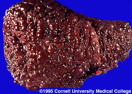
    · Some chronic carriers have chronic active hepatitis. This can lead to cirrhosis (the inflammation and hardening of the liver), hepatocellular carcinoma (primary liver cancer), and death.

http://www.pathology.med.ohio-state.edu/paxit/deptbase/Paxit/Images/10534/PAXIT025.JPG

http://www.stanford.edu/group/virus/hepadna/2004tansilvis/liver%20cirrhosis.gif

Laboratory Diagnosis


    · Quick detection for early HBV infection: immunoassay for HBsAg

    · HBsAg appears during the incubation period and is dewtectable in most patients during the acute disease. It falls to undetectable levels during convalescence in most cases

    · Prolonged presence of HBsAg indicates the carrier state and the risk of chronic hepatitis and hepatic carcinoma.

    · HBeAg is also detectable in acute infection which is characterized by a high rate of viral replication.

    · IgM antibodies against core antigen are detectable in serum.

    · Seubsequently, IgG antibodies against core are produced, and persist for life.

    · If the diagnosis of hepatitis B is confirmed, a prognosis may be assessed by liver biopsy.

Transmission

    · Through contaminated blood.

    · Sexual intercourse with an infected person. (horizontal transmission)

    · Perinatally from infected mother to newborn. (vertical transmission)

Epidemiology and Control

http://www.dshs.state.tx.us/idcu/disease/hepatitis/hepatitis_b/faqs/hepbdstb.gif

    · Estimated 400 million people worldwide infected with HBV.

    · Post 1987 babies all vaccinated against HBV.

    · Pre 1987 screening ongoing.

    · People at risk

o Doctors, laboratory workers, personnel coming in contact with blood.

o Drug addicts who share needles.

o Promiscuous behaviour.

Copyright © 2008 - The Virology Blog - is proudly powered by Blogger
Smashing Magazine - Design Disease - Blog and Web - Dilectio Blogger Template