Methods of Study of Viruses


Isolation and Cultivation of Viruses

Animals and eggs can be used for virus cultivation. However, this way of cultivating viruses has been mainly replaced by cell culture due to the inconvenience and safety involved in handling animals. Animals and eggs still have their advantages for use as a culture “media”—this method is still used to cultivate viruses that have no known host in vitro (e.g. influenza virus in chick embryo)), and to study viral pathogenesis in a whole host (e.g. polo studies in chimpanzees). Plants may also be used to cultivate certain plant viruses. The tobacco mosaic virus may be used to determine virus numbers based on the number of virus plaques on its leaves.

Cell tissue culture comprises of cells being grown in vitro on continuous cell lines. Continuous cells lines are the preferred choice of viral culture because they can be sub-cultured indefinitely, theoretically. They are derived from polyploid or multiploid cancerous cells. Viruses that do not grow in vitro have to be grown in animals, plants or eggs.

Detection, identification and diagnosis (elaborated later)

  1. Tissue culture methods

a. Cytopathic effect

b. Plaque assay

  2. Physical methods

a. X-ray crystallography

b. Electron microscopy

c. Ultracentrifugation

  3. Serological methods

a. Haemagglutination (HA)

b. Haemagglutination Inhibition (HI)

c. Virus neutralisation

d. Complement Fixation

  4. Immunological methods

a. Immunofluorescence

b. Immunogold EM

c. Immunoprecipitation

d. Immunoblot

e. Enzyme Linked Immunosorbent Assay (ELISA)

  5. Molecular biology methods to analyse viral proteins

a. SDS PAGE

b. Western blot

c. Protein sequencing

d. X-ray crystallography

  6. Molecular biology methods to analyse viral genome

a. Restriction analysis

b. DNA sequencing

c. Southern blot

d. Northern blot

e. PCR/RT-PCR

Cytopathic Effect:

Cytopathic effect (CPE) is an alteration in cell morphology resulting from viral infection of a cell culture monolayer. CPE may be used as a presumptive identification of a virus. If the virus does not produce a CPE, its presence can be detected by several other techniques such as haemadsorption.

http://www.microbelibrary.org/microbelibrary/files/ccImages/Articleimages/cummings/baculovirus.JPG

Plaque Assay:

Plaque Assays are used for the quantitative measure of infectious centres by counting the number of infectious virus particles in a given sample in Plaque forming units.

The plaque assay is based simply on the ability of a single infectious virus particle to give rise to a macroscopic area of cytopathology (i.e. plaque, focus of infection) on an otherwise normal monolayer of cultured cells.

Specifically, if a single cell in a monolayer is infected with a single virus particle, new virus resulting from the initial infection can infect surrounding cells which, in turn, produce virus that infects additional surrounding cells. Multiple rounds of infection give rise to an area of infection called a plaque.

*The semisolid medium prevents the formation of secondary plaques through diffusion of virus from the original site of the infection to new sites, ensuring that each plaque that develops in the assay originated from a single infectious particle in the starting inoculum.

http://www.v-bio.com/pictures/p06_small.png

Advantages

  1. Simple, inexpensive, precisely quantitative method.

  2. Higher accuracy at lower concentration—useful for samples with very low virus counts (in food and water samples).

Disadvantages/Limitations

  1. Counts only viable virus (virus capable of multiplying)

  2. Only works for viruses that infect monolayer cells and viruses that cause cell lysis (or other cytopathic changes that can be observed)

  3. Must know culture conditions for the virus studied.

  4. Requires time for incubation, time consuming.

Calculation

Original Virus Concentration (in PFU/ml) = Final Virus Concentration * Dilution Factor * (1/volume pipetted in well (in ml))

X-ray crystallography:

Viruses are crystallized and by using X-ray diffraction techniques, the structure of the virus can be elucidated from the diffraction pattern produced.

Electron microscopy:

This includes Transmission EM, Scanning EM and STEM. Electron microscopy involves scattering electrons on a specimen to “illuminate” it and magnify it for viewing. The resolution of an electron microscope is much greater than a light microscope as the wavelength of an electron is much smaller than that of a light proton.

http://www.biologie.uni-hamburg.de/b-online/ge03/14.gif

Ultracentrifuation:

Ultracentrifugation has assumed growing importance in virus diagnosis as a technique by which to concentrate and purify viruses for immediacy diagnosis on the basis of electron microscopy as well as for purely virological and serological tests.

Ultracentrifugation has proved to be helpful for sizeable improvement of sensitivity for detection, which, in turn, has been conducive to time saving. The preparational ultracentrifuge enables also direct diagnosis by determination of isodensities and sedimentation coefficients of viruses and their components.

Haemagglutination:

Agglutination of red blood cells caused by certain antibodies, virus particles or high molecular weight polysaccharides. This can be used to test for influenza and other viruses with two spike proteins neuraminidase and haemagglutinin, since these two proteins bind specifically to red blood cells.

Haemagglutination Inhibition:

This is used to quantify the virus by haemagglutination. If the virus and antibody are homologous, the antibody bound to the surface of the virus blocks its entry into the cell. This neutralizes infectivity, because it prevents viral replication and subsequent CPE formation or animal infection. Agglutination is also i

nhibited.


http://www.umanitoba.ca/science/microbiology/staff/cameron/graphics/401lab3passivehemagglutination%20group506.jpg

Virus neutralization:

If the virus and antibody are homologous, the antibody bound to the surface of the virus blocks its entry into the cell. This neutralizes viral infectivity, because it prevents viral replication and subsequent CPE formation or animal infection. It is one of the methods employed for the detection of virus-specific neutralizing antibodies.

Complement fixation:

Complement fixation is used to detect the presence of either specific antibody or specific antigen in a patient's serum. If the antigen (the unknown virus in the culture fluid) and the known antibody are homologous, complement will be fixed (bound) to the antigen-antibody complex. This makes it unavailable to lyse the “indicator” system, which is composed of sensitized red blood cells.

Immunofluorescence:

Microscopic method of determining the presence or location of an antigen (or antibody) by demonstrating fluorescence when the preparation is exposed to a fluorescein-tagged antibody (or antigen) using ultraviolet radiation.

http://www.microbelibrary.org/microbelibrary/files/ccImages/Articleimages/cummings/1245-MERGED%2020x%20wide%20field.JPG

Above: Immunofluorescence for Herpes Simplex Virus Antibody

Immunogold EM:

Locate specific proteins or antigens by attaching nano-gold particles to antibodies.

Immunoprecipitation:

Immunoprecipitation (IP) is the technique of precipitating a protein antigen out of solution using an antibody that specifically binds to that particular protein. This process can be used to isolate and concentrate a particular protein from a sample containing many thousands of different proteins. Immunoprecipitation requires that the antibody be coupled to a solid substrate at some point in the procedure.

Immunoblot:

The immunoblot (also called Western blot) is an analytical technique used to detect specific proteins in a given sample of tissue homogenate or extract. It uses gel electrophoresis to separate native or denatured proteins by the length of the polypeptide (denaturing conditions) or by the 3-D structure of the protein (native/ non-denaturing conditions). The proteins are then transferred to a membrane (typically nitrocellulose or PVDF), where they are probed using antibodies specific to the target protein.

ELISA (Enzyme-linked Immunosorbent Assay):

The basic principle of an ELISA is to use an enzyme to detect the binding of antigen (Ag) and antibodies (Ab). Antibodies are bonded to enzymes; the enzymes remain able to catalyze a reaction that converts a colourless substrate to a coloured product, indicating the presence of Ag:Ab binding. Depending on the test, an ELISA can be used to detect the presence of either Abs or Ags.

https://blogger.googleusercontent.com/img/b/R29vZ2xl/AVvXsEinBG2coFfuUX6aMFfIan1riPrk5KyKN8LrZiUjosvF0yIXlwJpVz4X9XDVZR9fLa9HUKOAl50exCkNaYjJvCW_udpAJqsFFTYu9600FRnzfsu6Vh1U-o3tTcu3fwdLKE6szx7qAzra5VAC/s1600-h/kakia.jpg

SDS PAGE:

SDS-PAGE, sodium dodecyl sulfate polyacrylamide gel electrophoresis, is a technique widely used in biochemistry, forensics, genetics and molecular biology to separate proteins according to their electrophoretic mobility.

Polymerase Chain reaction (PCR):

A method for expanding small discrete sections of DNA by binding DNA primers to sections at the ends of the DNA to be expanded and using cycles of heat (to create single-stranded DNA) and cooler temperatures (to allow a DNA polymerase enzyme to create new sections of DNA between the primer ends).

http://www.copernicusproject.ucr.edu/ssi/HighSchoolBioResources/Genetic%20Engin%20Hum%20Genome/pcr.jpg

Southern Blot:

Identification of specific genetic sequences by separating DNA fragments by gel electrophoresis and transferring them to membrane filters in situ. Labelled complementary DNA applied to the filter binds to homologous fragments, which can then be identified by detecting the presence of the labelled DNA in association with bands of certain molecular size. This test was named after its discoverer, E.M. Southern.

http://www.biogate.it/microarrays/images/Southern_blot.gif

Specific Viral Families: Poxviridae


Poxviridae

http://www.sciencedaily.com/images/2007/04/070430155718-large.jpg

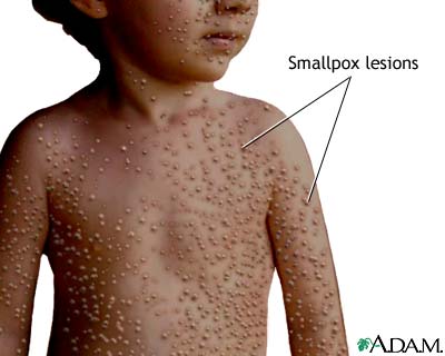
The poxviruses, another group of enveloped, linear dsDNA viruses, are the largest and most complex of all viruses. There are terminal repeated sequences and inverted repeats at both ends. They are widely distributed in nature. Nearly every animal species can be infected by a form of poxvirus. The human poxviruses (orthopoxviruses) are large, enveloped, brick-shaped viruses. These viruses multiply in specialized portions of the host cell cytoplasm called viroplasm, where they can cause skin lesions typical of smallpox and coxpox. Other poxviruses, such as monkeypox, can infect humans who have close contact with infected animals.

Important properties

    · Large family of viruses

    · Large enough just to be seen under a light microscope.

    · Unique oval-shaped

    · Antigenically very complex

    · Unlike the influenza virus, poxviruses can remain stable for hours in the air. It could be due to the fact that it is an enveloped virus.

Smallpox virus is an example of a poxvirus.

Transmission

    · Human is the only reservoir for smallpox

    · Respiratory secretions (aerosolized air droplets) and saliva and bodily fluids from infected person are sources of transmission

Clinical Features

    · ~12 days incubation

    · Initially influenza like symptoms

    · Characteristic pustules

    · Always associated with skin lesions and scarring of skins

    · Neurological damage

    · Blindness

    · Death

http://www.nlm.nih.gov/medlineplus/ency/images/ency/fullsize/19059.jpg

Two varieties of smallpox are:

1 Variola Major

a. 25%-30% fatalities.

  2 Variola Minor

a. Less than 1% death

Vaccination

In 1796, Edward Jenner developed a smallpox vaccine using cowpox. He used cowpox from milkmaids and inoculated it into an 8 year old boy James Phipps. When challenged with smallpox, he recovered quickly and gained immunity to smallpox.

Eradication of Smallpox

    · The variola virus has no reservoir except man

    · It only has a single, stable serotype, which is the key success of vaccination.

    · It causes only acute infection from which the patient dies, or

    · Obtains lifelong immunity to it as

    · Variola virus is an effective immunogen (sure to gain immunity from it)

    · Vaccination programs

    · Certified by WHO in 1980 to be defeated and eradicated off the face of the earth.

Laboratory Diagnosis

    · Diagnosis was made either by growing the virus in cell culture or chick embryos or by detecting viral antigens in vesicular fluid by immunofluorescence.

Control

    · Vaccination

    · Incineration of all infected material.

    · However, vaccination no longer required after 1980, when smallpox was eradicated.

    · Smallpox remains as one of the deadliest virus around—it is still kept in certain laboratories. There have been concerns that smallpox may be used in biowarfare and bioterrorism.

Iceberg Concept of Infection


Infection occurs

Specific Viral Families: Flaviviridae


Flaviviridae

The flaviviruses are enveloped, polyhedral, (+) sense RNA viruses that are transmitted by mosquitoes and ticks. The viruses produce a variety of encephalitides or fevers in humans. The yellow fever virus is a flavivirus that cause a haemorrhagic fever—in which blood vessels in the skin, mucous membranes and internal organs bleed uncontrollably. Hepatitis C infection is also caused by a flavivirus.

Important Properties

    · Spherical enveloped virion, 40-50nm

    · Inner core protein C

    · Membrane/matrix protein M

    · Envelope with glycoprotein peplomers (E)

    · Single linear 11kb positive sense ssRNA—infectious mRNA

    · Polyadenylated tail and 5’ cap

    · Cytoplasmic replication

    · Polyprotein from genomic RNA cleaved

    · 3 structural proteins

    · Several non-structural proteins

Dengue Virus

http://www.sciencedaily.com/images/2008/03/080327172348-large.jpg

  · Most important arbovirus presently.

    · Transmitted through the bite of a female Aedes aegypti mosquito (the vector).

    · 4 distinct serotypes based on neutralisation test.

    · DEN-1, DEN-2, DEN-3 and DEN-4

    · DEN-2 shows greatest antigenic and genotypic distance from the others.

    · Protective immunity after infection homotypic.


http://www.microbeworld.org/images/scientists/interviews/arbocycle.jpg

Dengue Infectious Cycle

Classic Dengue begins suddenly with an influenza syndrome consisting of fever, malaise, cough, and headache. Severe pains in muscles and joints occur. Enlarged lymph nodes, a masculopapular rash, and leukopenia are common. After a week or so, the symptoms regress but weakness may persist. Although unpleasant, this typical form of dengue is rarely fatal and has few sequelae.

Dengue Haemorrhagic Fever (DHF) is a much more severe disease, with a fatality rate that approaches 10%. The initial picture is the same as classic dengue, but then shock and haemorrhage, especially into the gastrointestinal tract and skin, develop. Dengue haemorrhagic fever occurs particularly in southern Asia, whereas the classical form is found in tropical areas worldwide.

Haemorrhagic Shock Syndrome (DSS) is due to the production of large amounts of cross-reacting antibody at the time of a second dengue infection. The pathogenesis is as dollows: The patient recovers from classic dengue caused by one of the four serotypes, and antibody against that serotype is produced. When the patient is infected with another serotype of dengue virus, an anamnestic, heterotypic response occurs, and large amounts of cross-reacting antibody to the first serotype are produced.

Pathogenesis of DHF/DSS

  1. Virulent strain theory

a. Some strains are more virulent than others

b. Molecular studies show variations in sequences amongst different strains within serotypes

c. Early evidence pointed to DEN-2

  2. Antibody enhancement

a. Main theory for DHF/DSS

b. Main cell target of DEN: Monocytes and macrophages

c. Most cases of DHF/DSS had prior infection or infants below 1 year had maternal Ab.

d. Monkey experiments showed similar enhancement.

Control of Dengue Virus

    · No antiviral therapy or vaccine for dengue is available.

    · Outbreaks are controlled by using insecticides and draining stagnant water that serves as the breeding ground for mosquitoes.

    · Personal protection includes using mosquito repellent and wearing clothing that covers the entire body.

http://www.stanford.edu/group/parasites/ParaSites2008/Nkem_Cristina%20Valdoinos/ugonabon_valdovinosc_dengueproposal_files/image002.png

Yellow Fever Virus

http://pathmicro.med.sc.edu/mhunt/yellow%20fever%20virus.jpg

  · Type species of the genus flavivirus

    · Tropical disease in Latin America and Africa

    · Transmitted through the bite of a female Aedes aegypti mosquito (the vector).

    · Incubation period: 3-6 days

    · Acute yellow fever—symptoms: Viremia, headache, malaise, nausea, muscle ache, flushing of head and neck, conjuntival infection, strawberry tongue.

    · Remission after acute yellow fever manifests as severe yellow fever—symptoms: fever, vomiting, abdominal pain, prostration.

    · Symptoms progress to involve the liver, kidneys and heart: e.g. jaundice.

http://lh4.ggpht.com/_YmfDLUdaIGU/SAFuz7jrw_I/AAAAAAAAAVM/4MOT98vdgxQ/Strawberry+tongue+%28Kawasaki+or+Scarlet+fever%29.jpg

Strawberry Tongue

Control of Yellow Fever Virus

    · Mosquito control (similar to dengue)

    · Immunnization with the vaccine containing live, attenuated yellow fever virus. Travelers to and residents of endemic areas should be immunized.

    · Protection lasts for up to 10 years; booster shots to be taken after that.

    · Because it is a live vaccine, it should not be given to immunocompromised people or to pregnant women.

West Nile Virus

  · Originated in Uganda

· Discovered in 1937

Symptoms

    · Usually mild to no symptoms

o Fever

o Headache, body aches

o Skin rash

o Swollen lymph glands

    · Severe Symptoms (occurring mainly in persons above 50 years old)

o Crossing blood-brain-barrier

o Encephalitis

o Meningitis

Transmission

    · Wild birds are the main reservoir of this virus

    · Vector is the mosquito, especially the Culex species

    · Humans are dead-end hosts

http://www.cdc.gov/ncidod/dvbid/westnile/misc/slides/komar/images/wnv-transmission-cycle.jpg

West Nile Virus Transmission Cycle

Control

    · No vaccine is available for west nile virus

    · Mosquito control

Specific Viral Families: Retroviridae


Retroviridae

The retroviruses are enveloped viruses that have two complete copies of (+) sense RNA. They also contain the enzyme reverse transcriptase which uses the viral RNA to form a complementary strand of DNA, which is then replicated to form a dsDNA. For the genetic information to be transcribed and translated, this DNA migrates to the host cell nucleus and become incorporated into chromosomes of host cells. Such integrated viral DNA is known as a provirus.

http://pathmicro.med.sc.edu/lecture/RNA2.jpg

Retroviruses cause tumours and leukemia in humans. The human retroviruses invade immune defense cells called T lymphocytes and are referred to as Human T cell Leukemia Viruses (HTLV). Both HTLV-1 and HTLV-2 are associated with malignancies (leukemia and other tumours), whereas the human immunodeficiency virus (HIV) causes acquired immune deficiency syndrome (AIDS).

Important Properties

    · Spherical enveloped virion, 80-100nm

    · Ribonucleoprotein in central nucleoid (concentric or truncated cone in lentivurses) within icosahedral capsid.

    · Envelope with glycoprotein peplomers

    · 2 copies of linear plus sense ssRNA each 7-10kb

    · 3’ polyadenylated tail and 5’ cap

    · Reverse transcriptase

    · Formation of long terminal repeats before prpovirus DNA inserted into host genome

    · Genome consists of gag, pol, env genes some regulatory genes, some oncogenes.

Human Immunodeficiency Virus

HIV is one of the human T-cell lymphotropic retroviruses. HIV preferentially infects and kills helper (CD4) T lymphocytes, resulting in a loss of cell mediated immunity and a high probability that the host will develop opportunistic infections. Other cells (e.g. macrophages and monocytes) that have CD4 proteins on their surfaces can be infected also.

https://blogger.googleusercontent.com/img/b/R29vZ2xl/AVvXsEhuSD_M4_xi0Hr1WcOR7MVcKfGUBOjidoN8TQMWhyphenhyphencbizC-ehgadoZnvoMqDJ1b4rauaIlZmedd4GIWq17Ah6UgpiqOVe9-wHK3nVKkZiFZfZhBUp89-23d0kfHIY7_oxrc0nZvkCsDLVpQ/s1600-h/aaaaad.jpg

Genes and Proteins of the Human Immunodeficiency Virus

Gene

Proteins Encoded by Gene

Function of Proteins

I. Structural Genes Found in All Retroviruses

gag

p24, p7

p17

Nucleocapsid

Matrix

pol

Reverse Transciptase

Protease

Integrase

Transcribes RNA genome into DNA

Cleaves precursor polypeptide

Integrates viral DNA into host cell DNA

env

gp120

gp41

Attachment to CD4 protein

Fusion with host cell

II. Regulatory Genes Found in Human Immunodeficiency Virus that are Required for Replication

tat

Tat

Activation of transcription of viral genes

rev

Rev

Transport of late mRNAs from nucleus to cytoplasm

III. Regulatory Genes Found in Human Immunodeficiency Virus that are NOT Required for Replication (accessory genes)

nef

Nef

Decreases CD4 proteins and class I MHC proteins on surface of infected cells; induces death of uninfected cytotoxic T cells; important for pathogenesis by SIV

vif

Vif

Stabilizes newly synthesized viral DNA and transports core through cytoplasm

vpr

Vpr

Transports viral core from cytoplasm into nucleus in non-dividng cells

vpu

Vpu

Enhances virion release from cell

Replicative Cycle of HIV

The replication of HIV follows the typical retroviral cycle.

  1. Inital step in the entry of HIV into the cell is the binding of the virion gp120 envelope protein to the CD4 protein on the cell surface.

  2. The virion gp120 protein then interacts with a second protein on the cell surface, one of the chemokine receptors (CXCR4 and CCR5). Individuals with mutations in the gene encoding CCR5 have protection from infection with HIV.

  3. The virion gp42 protein mediates fusion of the viral envelope with the cell membrane, and the virion enters the cell.

  4. After uncoating, the virion RNA-dependent DNA polymerase transcribes the genome RNA into double-stranded DNA, which integrates into the host cell DNA, mediated by integrase.

  5. The viral DNA can integrate at different sites in the host cell DNA, and multiple copies of viral DNA can integrate.

  6. Viral mRNA is transcribed from the proviral DNA by host cell ENA polymerase and translated into several large polyproteins.

  7. Precursor polyproteins can be assembled into nucleocapsid.

  8. As immature virions buds from the cell membrane, the polyproteins are cleaved by the viral protease. This results in the formation of mature, infectious virions.


http://www.nature.com/embor/journal/v4/n6s/images/embor857-f2.gif

Transmission

    · Transfer of infected blood.

    · Sexual contact (horizontal transfer)

    · Perinatal transmission from infected mother to neonate: Either across the placenta, at birth, or via breast milk. (vertical transfer)

Pathogenesis

http://www.nature.com/nri/journal/v3/n4/images/nri1058-f3.gif

  · Primary Infection

o Acute stage

o Flu-like symptoms

o Fever

o Skin rash

o Swollen skin nodes

o Due to virulence factors such as rate of replication, propensity to mutate and cytopathogenicity.

o Also due to host resistance mechanisms such as the suppression by CD8 T suppressor cells and the presence of cytotoxic T-lymphocytes.

    · Asymptomatic Stage

o No apparent disease

o Fall in CD4 T lymphocytes

o Possible signs:

§ Fatigue

§ Depression

§ Weight loss

§ Memory disorders

    · Symptomatic Stage

o AIDS-related complex

§ Diseases not considered definitive of AIDS

§ May be attributed to HIV infection

§ Indicative of defect in cell-mediate immunity

o AIDS

§ Opportunistic infections as a result of fall in CD4 T lymphocytes. (e.g. Kaposi’s Sarcoma)


http://www.aegis.com/images/aids-ois1.gif

Treatment

    · Highly active antiretroviral therapy (HAART)

o Consists of two nucleoside inhibitors (zidovudine and lamivudine) and a protease inhibitor (indinavir)

o Effective in prolonging life, improving the quality of life, and reducing viral load but does not cure the chronic HIV infection i.e. a latent infection of CD4+ cells continues indefinitely.

o Such combination therapy able to combat resistance—which arise due to rapid mutations due to inefficiency of reverse transcriptase.

    · Non-specific therapeutic management

    · Immunomodulation

o Enhancement of immune system through treatment with interleukin-2 (still under study)

    · No vaccine for human use is available.

Video from youtube: embed:

Prevention

Taking measures to avoid exposure to the virus:

    · Using condoms

    · Not sharing needles

    · Discarding donated blood that is contaminated with HIV

Copyright © 2008 - The Virology Blog - is proudly powered by Blogger
Smashing Magazine - Design Disease - Blog and Web - Dilectio Blogger Template