Specific Viral Families: Herpesviridae


Herpesviridae


http://pathmicro.med.sc.edu/virol/herpes_simplex.jpg

Above: TEM of Herpes Simplex Virus

Herpesviruses are a leading cause of human viral disease, second only to influenza and cold viruses. They are relatively large, enveloped viruses with linear dsDNA. Herpesviruses are widely distributed in nature.

Important properties

    · Concentric virion with

o Inner core

o Icosahedral capsid

o Amorphous tegument

o Envelope ( glycoprotein)

    · Linear dsDNA

    · Three origin of replication (ORI)

http://pathmicro.med.sc.edu/mhunt/dna15.jpg

Source:

The herpesviruses are known for their ability to cause latent infections.

    · In cells infected with herpesviruses, the viral dsDNA can exist as a provirus.

    · Herpesviruses remain in host cells, usually neurons, for long periods and retain the ability to replicate.

    · For example, a child who has recovered from chickenpox (varicella) will still have the virus in a latent form.

    · Years or decades later, the virus may be reactivated as a result of stress and/or physical factors.

    · This adult disease, which is very painful, is called shingles (zoster).

    · 11 of more than 100 genes of the herpesvirus genome are known to be involved in latency.

Pathogenesis of Human Herpes Type 1 to 5

Human Herpes Type

Name

Target cell Type

Disease

Latency

Transmission

1

Herpes simplex-1 (HSV-1)

Mucoepithelia

Oral herpes, encephalitis

Neuron

Close contact

2

Herpes simplex-2 (HSV-2)

Mucoepithelia

Genital and neonatal herpes, meningoencephalitis

Neuron

Close contact usually sexual

3

Varicella Zoster virus (VZV)

Mucoepithelia

Chickenpox (varicella) and Zoster (shingles)

Neuron

Contact or respiratory route

4

Epstein-Barr Virus (EBV)

B lymphocyte, epithelia

Infectious mononucleosis and Burkitt’s lymphoma; linked to Hodgkin’s disease, B cell lymphomas and to nasopharyngeal cancer.

B lymphocytes

Saliva

5

Cytomegalovirus (CMV)

Epithelia, monocytes, lymphocytes

Acute febrile illness; infections in immunosuppressed patients, leading cause of birth defects.

Monocytes, lymphocytes and possibly others

Contact, blood transfusions, transplantation, congenital

Clinical Features

HSV-1: Typically, Herpes labialis—Cold scores; Blisters around the mouth which lasts for around 1 week or more. May also affect the eyes (keratoconjunctivitis).

http://pathmicro.med.sc.edu/virol/coldsore2.jpg

HSV-2: Genital herpes, which is characterized by blisters, burning sensation and discharge.

VZV: In varicella, fever and malaise occur. Lesions appear on the trunk and spreads to the head and extremities. This is dangerous in pregnant women and may affect the nervous system—Guillain Barre syndrome. In zoster, painful vesicles occur along the course of a sensory nerve of the head or trunk.

http://pathmicro.med.sc.edu/virol/chickenpox3.jpg

Laboratory Diagnosis

    · Virus culture

    · Enzyme-linked immunosorbent assays (ELISAs)

    · Blood test

    · Tzanck smear

    · PCR assay

Epidemiology

HSV-1: Almost 100% of the adult population due to kissing and close proximity.

HSV-2: Up to 20% of the U.S. population due to sexual contact.

VZV: Varicella is a highly contagious disease of childhood; more than 90% of people in the U.S. have antibody by age 10 years. Varicella occurs worldwide.

EBV: Almost 100% of the adult population due to kissing and close proximity.

Control

HSV-1, HSV-2 and EBV: Avoid kissing to prevent contact with vesicular lesions or ulcers, and refrain from risky sexual behaviour. For the Herpes Simplex Viruses, caesarean section is recommended for women who are at term and who have genital lesions or positive viral cultures.

VZV: Vaccination with live, attenuated VZV (e.g. Varivax), and avoiding infected people.

Specific Viral Families: Hepadnaviridae


Hepadnaviridae

    · Small, enveloped, dsDNA (partially ssDNA) viruses.

    · The name hepadna comes from the infection of the liver—hepatitis—by a DNA virus.

    · Hepadnaviruses can cause chronic liver infections in humans and other animals, including ducks. In humans the hepatitis B virus causes hepatitis B, which can progress to liver cancer.

Hepatitis B Virus


http://pathmicro.med.sc.edu/lecture/hepatitis.gif

Important properties

  · 42-nm enveloped virion

    · Icosahedral nucleocapsid core contains a partially double-stranded circular DNA genome.

    · Envelope contains a protein called the surface antigen (HBsAg).

    · Core antigen (HBcAg) and the e antigen (HBeAg) are both located in the nucleocapsid protein but are antigenically different.

    · Endogeneous DNA-dependent DNA polymerase within the core.

    · Use of overlapping reading frame (ORF).

    · RNA-dependent DNA synthesis during replicative cycle.

Pathogenesis

    · Acute or chronic liver infection depending on the age at infection.

    · 90% of neonates and 50% of young children become chronically infected.

    · Only about 5% to 10% of immune-competent adults infected with HBV develop chronic hepatitis B.

    · A chronic carrier is someone who has HBsAg persisting in their blood for at least 6 months.

    · A high rate of heptatocellular carcinoma occurs in chronic carriers.

Basic characteristics of Hepatitis B infection:

Incubation period

60-90 days*

Fatality rate

1%

Recovery Rate

90%

Rate of Chronic Infection

<10%**

*with a range of 45-120 days

**although this can approach 90% in babies infected with HBV at birth.


http://www.medscape.com/content/2004/00/47/14/471470/art-jvh471470.fig2.gif

Above: The natural history of Hepatitis B

Clinical Features

Acute

    · Loss of appetite, nausea, vomiting, fever, abdominal pain and jaundice

    · About 90% - 95% of adults recover without sequelae

    · 5% - 10% become chronically infected

Chronic

    · While some chronic carriers will show clinical symptoms, most are asymptomatic—

they show no symptoms and may show no abnormalities on laboratory testing but remain infectious.

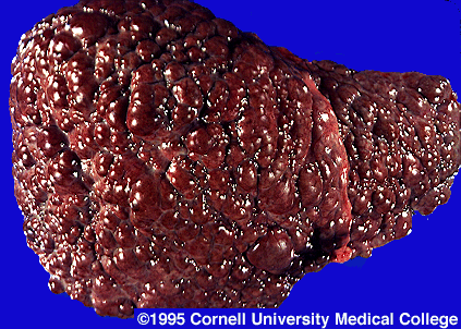
    · Some chronic carriers have chronic active hepatitis. This can lead to cirrhosis (the inflammation and hardening of the liver), hepatocellular carcinoma (primary liver cancer), and death.

http://www.pathology.med.ohio-state.edu/paxit/deptbase/Paxit/Images/10534/PAXIT025.JPG

http://www.stanford.edu/group/virus/hepadna/2004tansilvis/liver%20cirrhosis.gif

Laboratory Diagnosis


    · Quick detection for early HBV infection: immunoassay for HBsAg

    · HBsAg appears during the incubation period and is dewtectable in most patients during the acute disease. It falls to undetectable levels during convalescence in most cases

    · Prolonged presence of HBsAg indicates the carrier state and the risk of chronic hepatitis and hepatic carcinoma.

    · HBeAg is also detectable in acute infection which is characterized by a high rate of viral replication.

    · IgM antibodies against core antigen are detectable in serum.

    · Seubsequently, IgG antibodies against core are produced, and persist for life.

    · If the diagnosis of hepatitis B is confirmed, a prognosis may be assessed by liver biopsy.

Transmission

    · Through contaminated blood.

    · Sexual intercourse with an infected person. (horizontal transmission)

    · Perinatally from infected mother to newborn. (vertical transmission)

Epidemiology and Control

http://www.dshs.state.tx.us/idcu/disease/hepatitis/hepatitis_b/faqs/hepbdstb.gif

    · Estimated 400 million people worldwide infected with HBV.

    · Post 1987 babies all vaccinated against HBV.

    · Pre 1987 screening ongoing.

    · People at risk

o Doctors, laboratory workers, personnel coming in contact with blood.

o Drug addicts who share needles.

o Promiscuous behaviour.

Viral Classifications


Viral Classification

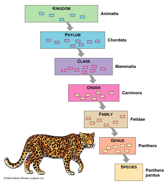
The usual means to classify and identify living things is to use the binomial system of naming devised by Carolus Linnaeus.

The hierarchy of classification:


http://www.mun.ca/biology/scarr/138599_KPCOFGS.jpg

One good mnemonic is: Keep Plates Clean Or Family Gets Sick!

However, because viruses are so different from cellular organisms, it is difficult to classify them according to the typical taxonomic categories (i.e. Kingdom, phylum). Instead, viruses may be classified with the ICTV classification, Lwoff’s scheme of classification, or the Baltimore’s system of classification.

ICTV Classification

    · Single, universal taxonomic scheme for viruses.

    · Devised by the International Committee on Taxonomy of viruses (ICTV).

    · Family is the highest taxonomic category used.

    · Criteria includes:

o Nature of genome & sequence relatedness

o Virus structure

o Natural host range

o Cell and tissue tropism

o Pathogenicity and cytopathology

    · Common name, rather than binomial term used to designate a viral species.

    · Problems such as distinguishing between viral species and viral strains have not been resolved completely.

Example:

Nidovirales (Order)àCoronaviridae (Family)àCoronavirus (Genus)àInfectious Bronchitis Virus (type species)

Note: A type species is a species whose name is linked to the use of a particular genus name. The genus is so typified will always contain the type species.

Lwoff’s Scheme of Classification

    · Devised in 1962 by Lwoff, RW Horne and P Tournier

    · Based on the physical properties of the virus:

o Nucleic acid

o Symmetry of capsid

o Presence/absence of envelope

o Dimensions of virion and capsid

Baltimore’s System of Classification

    · Devised by American biologist David Baltimore.

    · Encompasses all viruses

    · Based on the viral genome and its relationship to mRNA, modes of replication and gene expression.

    · Can make inferences and predictions about the fundamental nature of all viruses within each defined group.

    · Baltimore’s 1971 Paper titled “Expression of Animal Virus Genomes” : http://mmbr.asm.org/cgi/pmidlookup?view=long&pmid=4329869

There are 7 classes in the Baltimore Classification.

Class I (dsDNA)

Adenoviruses, Herpesviruses, Poxviruses, Papillomaviruses, Mimiviruses.

Class II (ssDNA)

Parvoviruses, Inoviruses, Microviruses, Anelloviruses, Nanoviruses.

Class III (dsRNA)

Reoviruses, Cystoviruses, Birnaviruses, Totiviruses, Partitiviruses.

Class IV ((+)-sense ssRNA)

Picornaviruses, Togaviruses, Flaviviruses, Astroviruses, Barnaviruses.

Class V ((-)-sense ssRNA)

Orthomyxoviruses, Paramyxoviruses, Rhabdoviruses, Filoviruses, Bunyaviruses.

Class VI (ssRNA reverse transcribing)

Retroviruses, Metaviruses, Pseudoviruses

Class VII (dsDNA reverse transcribing)

Hepadnaviruses, Caulimoviruses

General Characteristics of Viruses

General Characteristics of Viruses

Defining Viruses

  ·     Viruses are submicroscopic, acellular, obligate intracellular parasites—they replicate only inside a living host cell.

Components of Viruses

  ·     Viruses consist of a nucleic acid core and a protein capsid. Some viruses also have a membranous envelope.

  ·   Viral genetic information is contained in either DNA or RNA but never both.

  ·     Capsids are made up of subunits called capsomeres. They can have icosahedral symmetry, filamentous structure or complex structures.

  ·     A viral capsid and genome form a nucleocapsid. Such viruses are called naked viruses; those with a nucleocapsid surrounded by an envelope are enveloped viruses.

Sizes and Shapes of Viruses

  ·     Viruses have polyhedral, helical, binal, bullet or complex shapes and vary in size from 20 to 300nm in diameter.

Host Range and Specificity of Viruses

  ·     Viruses vary in host range and viral specificity. Many viruses infect a specific kind of cell in a single host species. Other viruses can infect several kinds of cells, several hosts, or both.

  ·     Viral specificity is determined by (1) whether a virus can attach to a cell, (2) whether appropriate host enzymes and other proteins the virus needs in order to replicate are available inside the cell, and (3) whether replicated viruses can be released from the cell to spread the infection to other cells.


Timeline of Virology


Timeline of Virology

·         1500 B.C.: Polio in Egypt

·         1200 B.C.: Smallpox in Egypt

·         11th century: Variolation practised in Asia and Africa: the deliberate infection of smallpox virus.

·         1796: Edward Jenner developed a smallpox vaccine using cowpox

·         1885: Louis Pasteur successfully developed rabies vaccination.

·         1892: Dimitri Ivanovski showed that the tobacco mosaic disease could be transmitted by extracts that were filtered by the filters fine enough to exclude the smallest known bacteria.

·         1898: Martinus Beijerinck found that the agent that caused the tobacco mosaic disease was not a mere toxin since it grew in the host.

·         1911: Francis Peyton Rous in 1911 described an oncovirus in chickens.

·         1911: Frederick Twort recognized the existence of viruses that infect bacteria.

·         1935: Wendell Stanley achieved the crystallization of the tobacco mosaic virus for electron microscopy and showed that it remains active even after crystallization.

·         1937: Max Delbruck described the basic life cycle of a virus.

·         1937: Max Theiler grew the Yellow Fever virus in chicken eggs and produced a vaccine from an attenuated virus strain.

·         1952: Hershey-Chase experiment showed that only DNA and not protein enters a bacterial cell upon infection with bacteriophage T2.

·         1955: Rosalind Franklin proposed the full structure of the tobacco mosaic virus.

·         1963: Hepatitis B virus discovered by Baruch Blumberg.

·         1965: Howard Temin described the first retrovirus.

·         1976: First recorded outbreak of Ebola hemorrhagic fever.

·         1977: Frederick Sanger achieved the first complete sequencing of the genome of any organism, the bacteriophage Phi X 174.

·         1977: Richard Roberts and Phillip Sharp showed that the genes of adenovirus contains introns and therefore require gene splicing.

·         1979: A world-wide vaccination campaign led by the UN World Health Organization resulted in the eradication of smallpox.

·         1982: Stanley Prusiner discovered prions and showed that they cause scrapie.

·         1985: Harald zur Hausen showed that two strains of the Human Papillomavirus (HPV) cause most cases of cervical cancer.

·         2002: Poliovirus was synthetically assembled in the laboratory.

·         2003: Bacteriophage Phi X 174 was synthetically assembled in the laboratory.

·         2004: Giant mimivirus was sequenced.

·         2006: Two vaccines protecting against the two cancer causing strains of the HPV were released.


Koch’s Postulates

Koch’s Postulates (a set of criteria published by Koch in 1890) provide a way to link a pathogen with a disease:

  1. A specific causative agent must be observed in every case of a disease.

  2. The agent must be isolated from a host displaying the disease and grown in pure culture.

  3. When the agent from the pure culture is inoculated into an experimentally healthy, susceptible host, the agent must cause the disease.

  4. The agent must be reisolated from the inoculated, diseased experimental host and identified as the original specific causative agent.

When Koch’s Postulates are met, an organism has been proved to be the causative agent of an infectious disease.

It is relatively easy to demonstrate that each postulate is met for bacterial diseases. However, it is more difficult to demonstrate the postulates in viral diseases since viruses cannot be grown in artificial media and must instead be grown in living cells. Furthermore, viruses are usually host specific. Thus, this should be noted—that not all infectious agents will grow in a given media, and the causative agent might not be detected.


https://blogger.googleusercontent.com/img/b/R29vZ2xl/AVvXsEiuUPOZg8ZXkboNAt_nUgqsYyXawnP7mGCaoTJVwJQCF1ATGboi0pR_PNmc9Wwvt2ZLbbrSP6ViMFoHII8uhrGeIq1rSBR8hssPvn__t7clIsH2xn94dF6V_5XAtwDIccimLZ5sg3r-3rqd/s1600-h/14-03_koch_1.jpg

Copyright © 2008 - The Virology Blog - is proudly powered by Blogger
Smashing Magazine - Design Disease - Blog and Web - Dilectio Blogger Template