Introduction!

This is a blog on General Virology. Virology is a branch of science that deals with viruses and viral diseases. All facts and data represented here have been summarised from the notes given to us by our helpful lecturers, and from the websites and books that we researched on.

This website was designed in a simple and concise manner, to provide you, the reader, with the knowledge you need to acquire.

All contents of the blog may be browsed using the archives links on the right hand side.

This is a project proudly done by:

Wong Wei Sheng Wilson (082571G)
Liang Weisong Edwin (083818Q)
Tan Jun Yan Eugene (083727K)
Nabila Binte Muhammad Nubli (084326D)
Nur Nurulshahqairani Binte Azmi (082673C)


Prions, Viroids and Virusoids


In the 1920s, several cases of a slow but progressively dementing illness in humans were observed independently by Hans Gerhard Creutzfedlt and Alfons Maria-Jakob. It was characterized by mental degeneration, loss of motor function, and eventual death.

In 1982, Stanley Prusiner proposed that the infective causative agent is an exceedingly small proteinaceous infectious particle called a prion.

Introducing Prions

Prions are rogue proteins that transform other cellular protein (PrPC) to the prion form PrPSC. Research indicates that prions are normal proteins that become folded incorrectly (possibly due to mutations).

Prions cause fatal, neurological degenerative diseases, including Creutzfeldt-Jakob disease (CJD), kuru, scrapie (Transmissible spongiform encephalopathy), mad cow disease (Bovine spongiform encephalopathy) and chronic wasting disease. The disease can be passed on to humans—new-variant CJD.

Important Characteristics

   ·      Prions are resistant to inactivation by heating to 90°C, which will inactivate viruses.

   ·       Prion infection is not sensitive to radiation treatment that damages virus genomes.

   ·       Prions are not destroyed by enzymes that digest DNA or RNA.

   ·       Prions are sensitive to protein denaturing agents such as phenol and urea.

   ·       Prions have direct pairing of amino acids.

Mechanism of Action

http://www.bio.davidson.edu/Courses/Molbio/MolStudents/spring2005/Winter/clip_image012.jpg

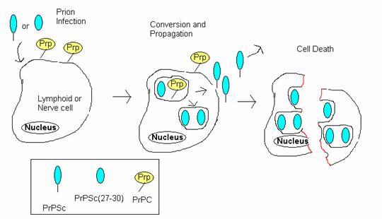
   1.      Initial infection and eventual interaction with normal cellular PrPC.

a.       PrPSc forms a heterodimer with normal PrPC

b.      Template for altering the protein fold

c.       Tightly coiled alpha helix becomes converted to loose beta sheets.

   2.      PrPC and PrPSc are internalized and interact within caveolae-like vesicles where PrPC is converted to PrPSc.

   3.     PrPSc may be recycled to the cell surface(shedding) to interact with PrPC from other cells and spread the infection, or remain and accumulate within the cell.

   4.      When PrPSc accumulates in the cell, they stick together inside cells, forming small fibrils. Because the fibrils cannot be organized in the plasma membrane correctly, such aggregations eventually kill the cell.

   5.      Cell death would release PrPSc and spread the infection; partially released proteolyzed PrPSc 27-30 may aggregate within plaques.

http://www.bio.davidson.edu/Courses/Molbio/MolStudents/spring2005/Winter/clip_image010.jpg

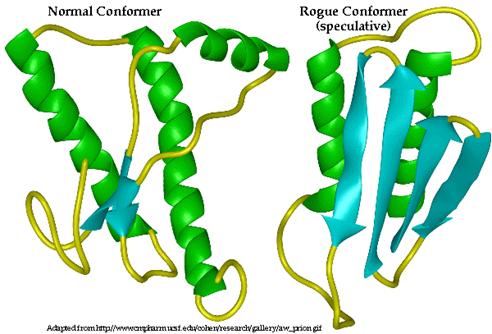
In Step 1(c): Three-dimensional structure of PrP C (left) and proposed 3D structure of PrP Sc (right). Alpha helices are indicated in green while beta sheets are indicated in blue. PrP C is composed primarily of alpha helices while PrP Sc is composed primarily of Beta pleated sheets.

Note:

·       α-helical, cellular isoform of the prion protein is PrPC. It is a membrane-anchored glycoprotein.

        ·       Insoluble, β-sheet, infectious, disease-causing isoform is PrPSc

PrP C

PrP Sc

primarily alpha helix structure

primarily beta sheet structure

protease susceptible

resistant to protease

monomeric species

forms multimeric aggregates

stable monomer

less stable monomer, self propagates for stability

normal resistance

extremely resistant to heat, chemicals, radiation and strong solvents


detergent soluble

detergent insoluble

 Evidence that PrP is the protein involved.

·         Mouse with knockout PrPC gene

·         No PrPC protein

·         Infect with prions

·         Nothing to convert

·         Mouse lives

Mouse with normal PrPC gene

Produce PrPC protein

Infect with prions

Prions convert normal PrPC to rogue proteins

Mouse dies


Pathogenesis

On the light-microscopic level spongiform changes in the brain, degeneration of neurons and astrocytosis are histopathological hallmarks of prion diseases.

Transmission:

Eating beef or mutton contaminated with prions.

Model of propagation of PrPSc

Viroids

   ·       Viroids are infectious agents composed exclusively of a single piece of circular single stranded RNA which has some double-stranded regions.

   ·       They mainly cause plant diseases (25 main sequences identified) by interfering with mRNA processing—they are catalytic ribozymes that cleave RNA to produce fragments containing a 5’-hydroxyl and a 2’, 3’-cyclic phosphate.

   ·       Only one sequence is known to infect man (Hepatitis D)

Hepatitis D

   ·       Only human disease known to be caused by a viroid.

   ·       Previously called delta agent

   ·       There is extensive sequence complementarity between the hepatitis D viroid RNA and human liver cell 7S RNA

o   The 7S RNA structure involved in the translocation of secretory and membrane-associated particles.

   ·         Hepatitis D viroid causes liver cell death via sequestering this 7S RNA and/or cleaving it.

Transmission

   ·       Co-infection with Hepatitis B virus

   ·       Bodily fluids

o   Unprotected sex

o   Sharing contaminated needles

o   Close proximity

http://www.thailabonline.com/blood/hepdslide_1.gif

Virusoid

Virusoids are infectious agents that infect plants in conjunction with an assistant virus.

They are not considered viruses but are subviral particles.

The size and structure is similar to viroids.

Its genome is a single molecule of single stranded circular RNA that is several hundred nucleotides long and codes for nothing but its own structure.

Emerging Viruses


Definition of emerging viruses:

Known viruses that have newly appeared in a human population and is rapidly increasing in disease incidence.

Source of emerging viruses:

    · Animals are usually the reservoirs of emerging viruses (zoonotic viruses).

    · Viruses were probably endemic at low levels in localized areas.

    · However, they could have “jumped” species, acquired a new host range and spread from there.

Examples of emerging viruses:

Dengue virus, Avian Influenza virus (e.g. H5N1 strain), SARS coronavirus, Nipah virus and Ebola virus.

http://bepast.org/docs/photos/nipah%20virus/nipah%20virus.jpg

Nipah Virus

http://m.blog.hu/li/lightscience/image/gpw-20050430a-fullsize-Ebola-virus-CDC-PHIL-ID-1181.jpg

Ebola Virus

http://ec.europa.eu/health/ph_threats/com/Influenza/images/influenza.jpg

Reasons for Emergence

Virus Factors

    · Spontaneous evolution of a new virus entity

    · Generation of a novel strain due to co-infection of different strains in an individual (random assortment)

Human Factors

    · Concentration of people with shared lifestyle

o E.g. the sharing of hypodermic needles, unsafe sex

    · Breakdown in public health

o Mainly in third world countries

    · Climate change

o E.g. increased wet seasons will lead to more stagnant water which might make it more conducive for mosquito breeding in a particular area. Aedes mosquito is a vector of dengue fever. Thus, the spread of the dengue flavivirus will be enhanced.

    · Man invading natural habitat of animal

o Deforestation for agricultural needs

o Closer to wild animals

o Increasing the possibility of zoonoses

General Viral Replication Cycle

Viral Growth Curve

· The amount of virus is plotted on a logarithmic scale as a function of time after infection.

· At the start, the virus disappears but viral nucleic acid continues to function and begins to accumulate within the cell as the virus is replicating in the cell. The time during which no virus is found inside the cell is known as the eclipse period. The eclipse period ends with the appearance of virus.

· The latent period is defined as the time from the onset of infection to the appearance of virus extracellularly.

· Alterations of cell morphology accompanied by marked derangement of cell function begin toward the end of the latent period.

· At the end of the latent period, new progeny viruses are assembled and released as the cell bursts.

· Infection begins with one virus particle and ends with several hundred virus particles having been produced. This is known as productive infection.

Viral Growth Cycle

6 main steps:

  1. Attachment

  2. Entry (penetration)

  3. Uncoating

  4. Replication of genome and gene expression

  5. Assembly

  6. Release

http://staff.vbi.vt.edu/pathport/pathinfo_images/CCHFV/rc.jpe

Attachment

    · The virus attachment proteins on the surface of the virion attach to specific receptor proteins on the host cell surface through weak, noncovalent bonding.

    · The specificity of attachment determines the host range of the virus.

    · Receptor on host cell mediates viral entry into cell.

Routes of Host Entry

    · Clathrin coated pits in endocytosis: Naked viruses such as the Hepatitis C virus.

    · Receptor-mediated endocytosis: Enveloped viruses such as the Epstein Barr Virus.

    · Fusion: Lipid bilayer of virus fuses with lipid bilayer of host cell.

    · Direct penetration: Translocation of entire virus particle mediated by receptor.

Uncoating

    · The viral genome must be separated from its protein coat once the virus enters the host cell’s cytoplasm. This process is called uncoating.

    · Naked viruses are uncoated by proteolytic enzymes from host cells or from the viruses themselves.

    · The uncoating of viruses such as the poxviruses is completed by a specific enzyme that is encoded by viral DNA and formed soon after infection.

    · Polioviruses begin uncoating even before penetration is complete.

Replication and Expression

    · After uncoating has taken place, genome replication and gene expression takes place—mRNA is synthesized, processed and translated to produce viral proteins.

    · At this point, viruses follow different pathways depending on the nature of their nucleic acid and the part of the cell in which they replicate.

Assembly

    · Assembly depends on the site of synthesis

    · Sites of protein synthesis and processing are the endoplasmic reticulum and Golgi body.

    · Sites for assembly are the nucleus, endoplasmic reticulum and Golgi body.

    · The progeny particles are assembled by packaging the viral nucleic acid within the capsid proteins.

Release

    · Virus buds off the plasma membrane of the host cell, and an envelope forms around the new viruses.

o Virus specific proteins enter the cell membrane at specific sites

o The viral nucelocapsid then interacts with the specific membrane site mediated by the matrix protein.

o The cell membrane evaginates at that site, and an enveloped particle buds off from the membrane.

o This usually occurs with enveloped viruses. An exception is the herpesviridae family, whose members acquire their envelopes from the nuclear membrane rather than the outer cell plasma membrane.

    · Alternatively, there may be an accumulation of particles in vesicles and release via exocytosis. This usually occurs with enveloped viruses.

    · Host cell lysis may follow.


Life Cycle of HIV virus

Copyright © 2008 - The Virology Blog - is proudly powered by Blogger
Smashing Magazine - Design Disease - Blog and Web - Dilectio Blogger Template